
Our Services

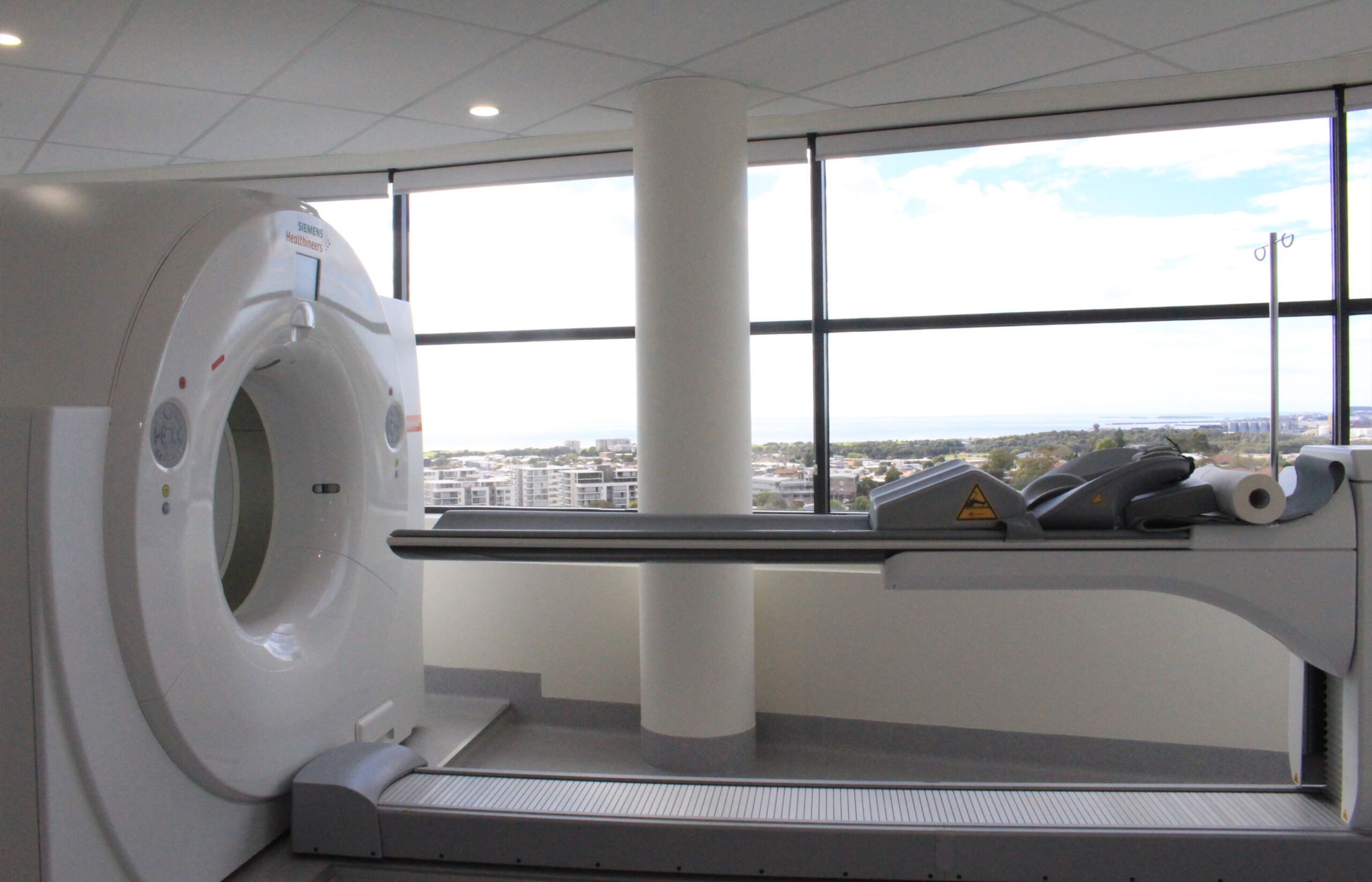
When you visit our clinic for a radiological study, you can enjoy district views with comfortable window seating in our observation room.
PET-CT (FDG & PSMA)

Siemens Biograph mCT(64)-3R (64 Slice CT) PET-CT

This brand new hybrid machine offers both PET and CT, meaning you can have all your scans performed in the one location. It features a powerful CT solution with 64-slice imaging, for enhanced detectability of lesions, as well as a 78cm wide imaging centre for superior patient comfort. 

A PET-CT, or Positron Emission Tomography-Computed Tomography, is a medical imaging technique that combines two types of imaging technologies: Positron Emission Tomography (PET) and Computed Tomography (CT). It is used in medicine to diagnose, stage, and monitor various diseases, most commonly cancer.

Here’s how a PET-CT works:

  1. Positron Emission Tomography (PET): In PET imaging, a small amount of a radioactive tracer, usually a type of sugar called fluorodeoxyglucose (FDG), is injected into the patient’s bloodstream. Cancer cells and some other abnormal cells take up this radioactive tracer at a higher rate than normal cells because they are more metabolically active. As the tracer decays, it emits positively charged particles called positrons.
  2. Detection of Positrons: Special detectors called gamma cameras or PET scanners detect the positrons emitted by the tracer. When a positron encounters an electron within the body, they annihilate each other, emitting gamma rays in opposite directions.
  3. Image Reconstruction: The PET scanner collects information about the distribution of the radioactive tracer within the body. This data is then used to create a three-dimensional image that shows the areas of high tracer uptake. These areas are often indicative of disease, such as cancer or areas of inflammation.
  4. Computed Tomography (CT): In a CT scan, X-rays are used to obtain detailed cross-sectional images of the body’s anatomy. CT scans provide information about the structure and location of abnormalities.
  5. Fusion of Images: The PET and CT images are superimposed or “fused” to create a single combined image. This fusion allows doctors to precisely pinpoint areas of increased metabolic activity (from the PET scan) within the context of the patient’s anatomy (from the CT scan).


The PET-CT scan provides valuable information to healthcare professionals, helping them determine the extent and location of diseases, assess the response to treatment, plan surgeries, and guide radiation therapy. It is commonly used in oncology for cancer staging, but it can also be used in cardiology, neurology, and other medical specialties to diagnose and monitor various conditions.

PET-CT studies we offer:

SPECT-CT (General Nuclear Medicine)

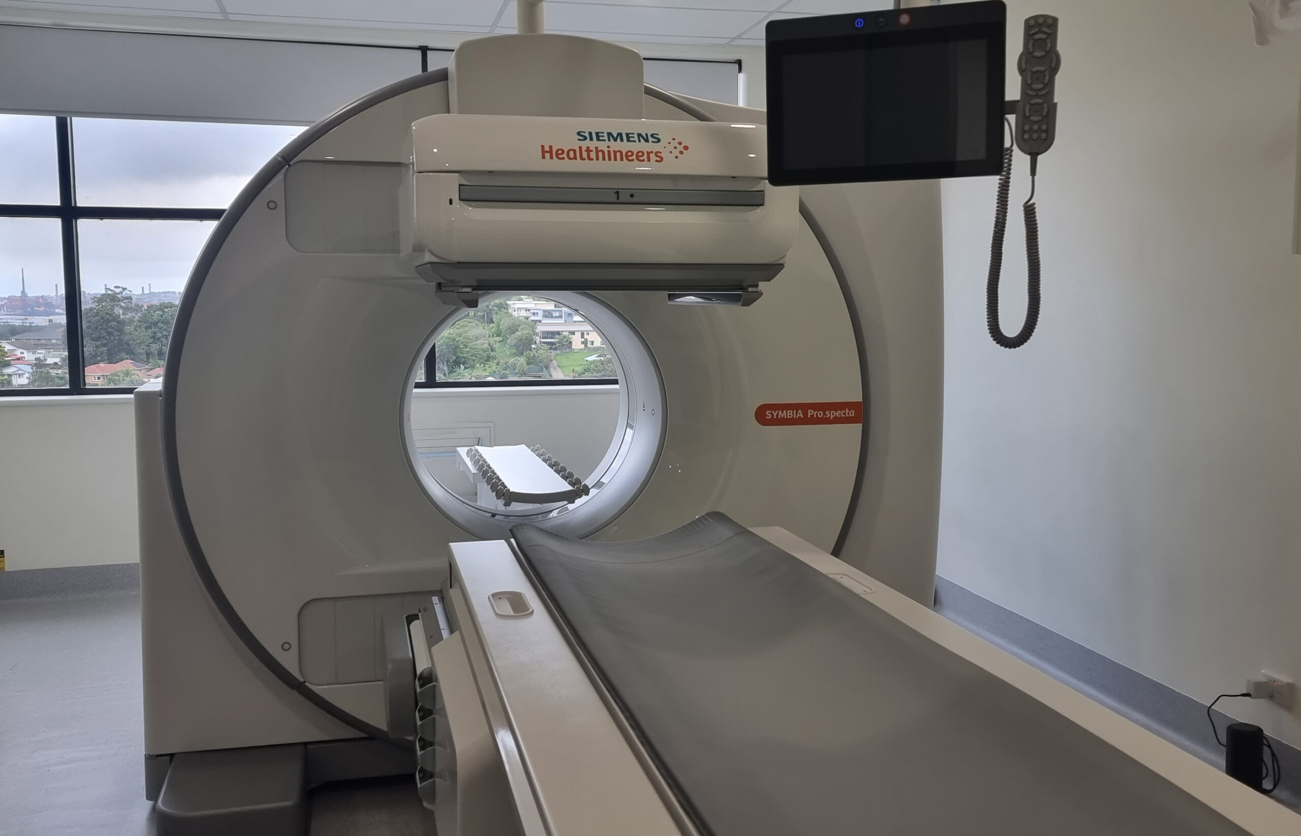
Siemens Symbia Pro.specta SPECT-CT

Released to market in Sep/Oct 2023, this brand new cutting edge machine is currently the newest in Australia (as at Jan 2024) featuring the latest advanced software and low-dose tin filter radiation reducing technology.

A SPECT/CT, or Single Photon Emission Computed Tomography/Computed Tomography, is a medical imaging technique that combines two imaging modalities: SPECT and CT. This hybrid imaging technology is used primarily for diagnosing and evaluating various medical conditions, particularly in the fields of nuclear medicine and radiology.


Here’s how SPECT/CT works:

  1. Single Photon Emission Computed Tomography (SPECT): SPECT is a nuclear medicine imaging technique that involves the use of a radioactive tracer or radiopharmaceutical. This tracer is injected into the patient’s bloodstream, and it accumulates in specific organs or tissues of interest, depending on the medical condition being studied. The radiopharmaceutical emits gamma rays, which are detected by a gamma camera.
  2. Gamma Camera Imaging: A gamma camera rotates around the patient and captures images of the gamma rays emitted by the radiopharmaceutical. These images provide information about the distribution and intensity of the radioactive tracer within the body. SPECT allows for functional imaging, showing the physiological processes in the body, such as blood flow, organ function, or tumor metabolism.
  3. Computed Tomography (CT): In a CT scan, X-rays are used to create detailed cross-sectional images of the body’s anatomy. CT scans provide high-resolution structural information about the body’s internal organs and tissues.
  4. Fusion of Images: The SPECT and CT images are fused or overlaid to create a single, combined image. This fusion allows healthcare professionals to precisely correlate the functional information from the SPECT scan with the anatomical details from the CT scan. It helps in localising abnormalities seen on SPECT images within the context of the patient’s anatomy.


SPECT/CT is used in various clinical applications, including:

  1. Oncology: It aids in the staging, restaging, and monitoring of cancer by combining functional information (SPECT) with anatomical information (CT).
  2. Cardiology: SPECT/CT can assess myocardial perfusion, evaluate cardiac function, and detect areas of reduced blood flow in the heart.
  3. Neurology: It can help in diagnosing neurological disorders, such as epilepsy or brain tumors, by providing functional and structural information.
  4. Orthopedics: SPECT/CT is used to assess bone health, detect fractures, and evaluate joint and bone diseases.
  5. Infection imaging: It can help locate areas of infection or inflammation in the body.


The combination of SPECT and CT in a single imaging session provides a comprehensive view of a patient’s condition, making it a valuable tool for diagnosing and managing a wide range of medical conditions.
SPECT-CT studies we offer:

DEXA X-Ray

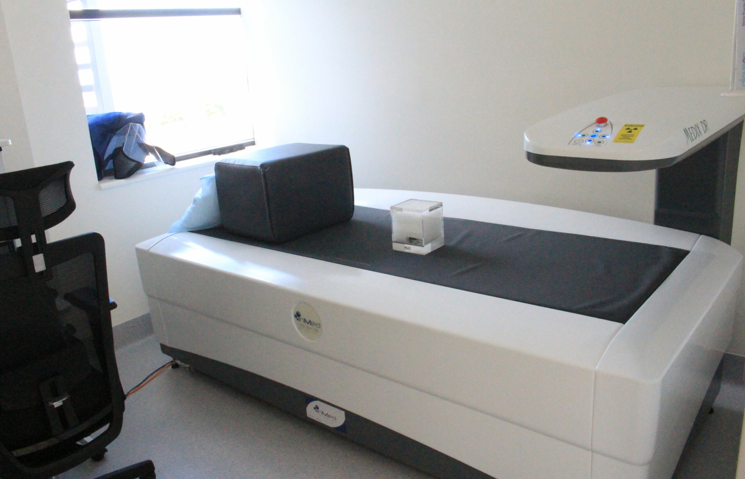
Medilink Medix DR Narrow Angle Fan Beam DEXA X-Ray

This brand new machine uses Narrow Angle Fan Beam Technology which results in 84-times lower radiation dose for whole body scans, and up to 5-times lower radiation dose for hip scans, when compared to wide angle fan beam technology found elsewhere.

A DEXA X-ray, also known as Dual-Energy X-ray Absorptiometry, is a medical imaging technique used to measure bone density and assess the risk of osteoporosis or bone-related conditions. DEXA scans are non-invasive and are commonly used to evaluate bone health, especially in postmenopausal women and older adults.

Here’s how a DEXA scan works:

  1. X-ray Beams: During a DEXA scan, two different X-ray beams, typically of differing energy levels, are directed at the bones being examined.
  2. X-ray Absorption: The X-ray beams are absorbed differently by bone and soft tissue. Bones are denser and absorb more X-rays, while soft tissues allow more X-rays to pass through.
  3. Detector Measurement: Detectors positioned on the opposite side of the X-ray source measure how much of each X-ray beam is absorbed by the bone and soft tissue.
  4. Bone Density Calculation: The data from the detectors are used to calculate bone density at specific locations, typically the hip, spine, and sometimes the forearm. These measurements are often expressed as grams of mineral per square centimeter.
  5. Comparison to Reference Data: The results are compared to a reference database of bone density values for individuals of the same age, sex, and ethnicity. A T-score is calculated, which quantifies how a person’s bone density compares to that of a healthy young adult. A Z-score may also be provided, which compares the patient’s bone density to individuals of the same age.


DEXA scans are useful for several purposes:

  • Osteoporosis Diagnosis: DEXA scans can help diagnose osteoporosis by assessing bone density. Osteoporosis is characterized by low bone density and increased susceptibility to fractures.
  • Fracture Risk Assessment: They can assess an individual’s risk of fractures, which can help in preventive measures and treatment decisions.
  • Monitoring Treatment: DEXA scans are used to monitor the effectiveness of osteoporosis treatments and interventions over time.
  • Research and Baseline Data: DEXA scans are also used in research studies and clinical trials to collect baseline data on bone density and assess the impact of interventions.


DEXA scans are safe, quick, and relatively low in radiation exposure compared to traditional X-rays. They play a crucial role in managing bone health and helping healthcare professionals make informed decisions regarding bone-related conditions and treatment plans.

DEXA studies we offer:

Get in touch today!

Speak with one of our friendly team members to book an appointment.