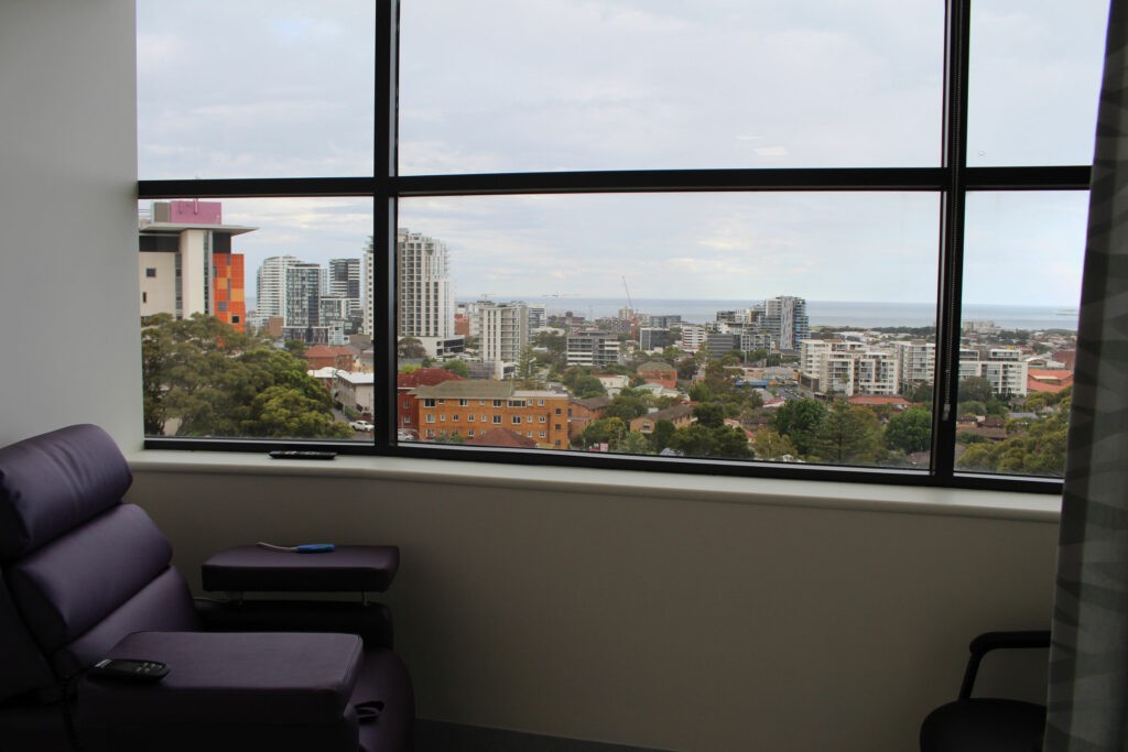
When you visit our clinic for a radiological study, you can enjoy district views with comfortable window seating in our observation room.
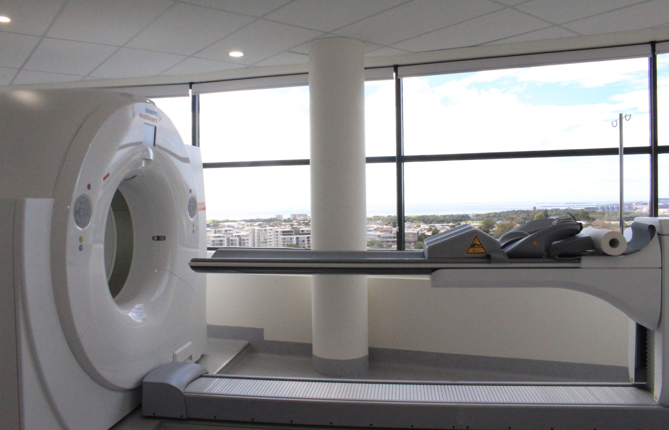
This brand new hybrid machine offers both PET and CT, meaning you can have all your scans performed in the one location. It features a powerful CT solution with 64-slice imaging, for enhanced detectability of lesions, as well as a 78cm wide imaging centre for superior patient comfort.
A PET-CT, or Positron Emission Tomography-Computed Tomography, is a medical imaging technique that combines two types of imaging technologies: Positron Emission Tomography (PET) and Computed Tomography (CT). It is used in medicine to diagnose, stage, and monitor various diseases, most commonly cancer.
Here’s how a PET-CT works:
The PET-CT scan provides valuable information to healthcare professionals, helping them determine the extent and location of diseases, assess the response to treatment, plan surgeries, and guide radiation therapy. It is commonly used in oncology for cancer staging, but it can also be used in cardiology, neurology, and other medical specialties to diagnose and monitor various conditions.
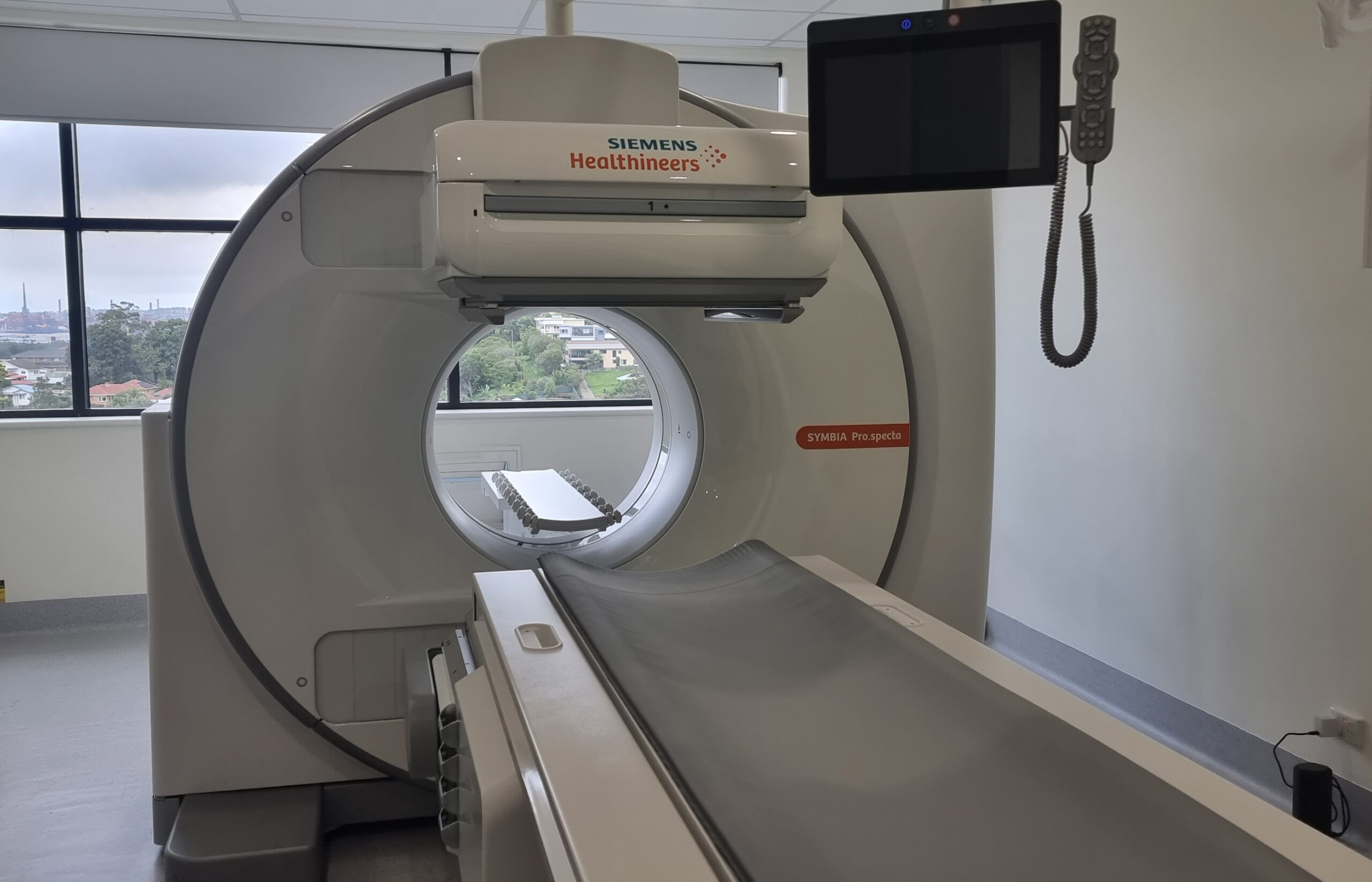
Released to market in Sep/Oct 2023, this brand new cutting edge machine is currently the newest in Australia (as at Jan 2024) featuring the latest advanced software and low-dose tin filter radiation reducing technology.
A SPECT/CT, or Single Photon Emission Computed Tomography/Computed Tomography, is a medical imaging technique that combines two imaging modalities: SPECT and CT. This hybrid imaging technology is used primarily for diagnosing and evaluating various medical conditions, particularly in the fields of nuclear medicine and radiology.
Here’s how SPECT/CT works:
SPECT/CT is used in various clinical applications, including:
The combination of SPECT and CT in a single imaging session provides a comprehensive view of a patient’s condition, making it a valuable tool for diagnosing and managing a wide range of medical conditions.
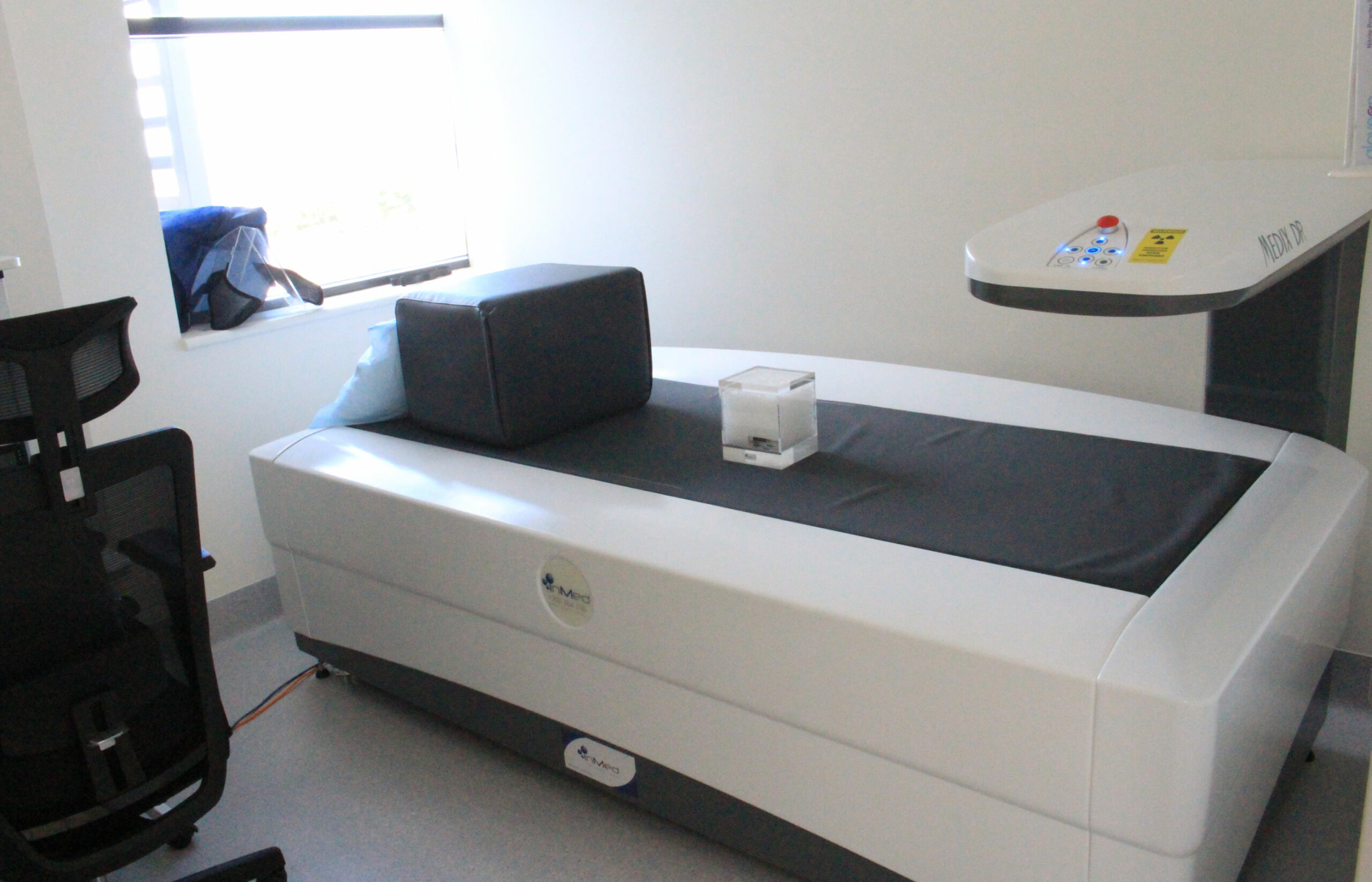
This brand new machine uses Narrow Angle Fan Beam Technology which results in 84-times lower radiation dose for whole body scans, and up to 5-times lower radiation dose for hip scans, when compared to wide angle fan beam technology found elsewhere.
A DEXA X-ray, also known as Dual-Energy X-ray Absorptiometry, is a medical imaging technique used to measure bone density and assess the risk of osteoporosis or bone-related conditions. DEXA scans are non-invasive and are commonly used to evaluate bone health, especially in postmenopausal women and older adults.
Here’s how a DEXA scan works:
DEXA scans are useful for several purposes:
DEXA scans are safe, quick, and relatively low in radiation exposure compared to traditional X-rays. They play a crucial role in managing bone health and helping healthcare professionals make informed decisions regarding bone-related conditions and treatment plans.